Cell and Tissue Imaging Unit
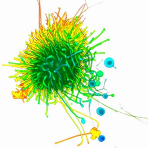
Our imaging core facility provides the instruments to image biological processes from tissues to single cells and subcellular molecular kinetics with both conventional and high-throughput methods.
Leaders
Contact persons
EQUIPMENT
Equipment includes Opera Phenix Plus high content confocal microscope system, Zeiss LSM 700 and LSM 800 Airyscan confocal microscopes, ISS M612 TCSPC/FLIM confocal microscope, Incucyte S3 and SX5 high-content imaging systems for living cells, Leica THUNDER Imager 3D Tissue slide scanner, Evident BX63 wide-field fluorescence microscope, and an image processing computer with Imaris 10.
LOCATION
- Opera Phenix Plus is located in Snellmania Building, 3rd floor, room SN3153.
- Confocal microscopes, TCSPC/FLIM microscope and Leica Thunder slide scanner is located in Snellmania Building, 3rd floor, room SN3151.
- Evident BX63 wide-field fluorescence microscope is located in Snellmania Building, 3rd floor, room SN3155.
- Image analysis computer with Imaris is located in Snellmania Building, 3rd floor, room SN3155.
- Incucyte S3 and SX5 imaging platforms are located in Snellmania Building, 3rd floor, room SN3156.
- Histology laboratory is located in Snellmania Building, 3rd floor, room SN3245.
NB! The doors to the imaging unit premises are locked 24/7. You can apply for a key through your unit’s or department’s key responsible person.
CONTACT
Janne Capra (Core Manager), janne.capra(at)uef.fi, p. 050-5165268, room SN3209.
Keywords
Leaders
Senior Researchers
-

Hanna Grobe
Staff ScientistInstitute of Biomedicine, School of Medicine, Faculty of Health Sciences
Post-doctoral Researchers
Technicians
-
Eija Rahunen
Senior Laboratory TechnicianInstitute of Biomedicine, School of Medicine, Faculty of Health Sciences -
Minna Turunen
BioanalystInstitute of Biomedicine, School of Medicine, Faculty of Health Sciences


