Multiscale Imaging Group
Research group
01.09.2015 -
A.I. Virtanen Institute for Molecular Sciences, Faculty of Health Sciences
Leaders
Contact persons
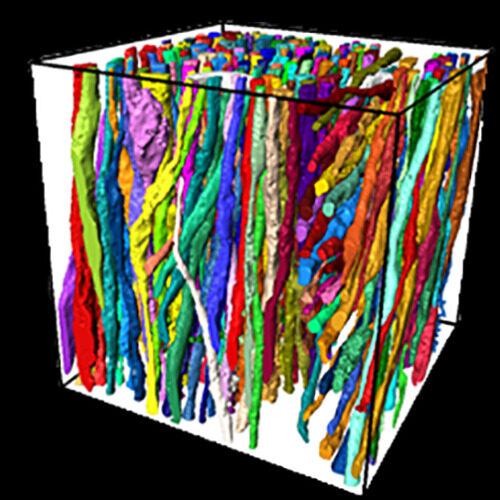
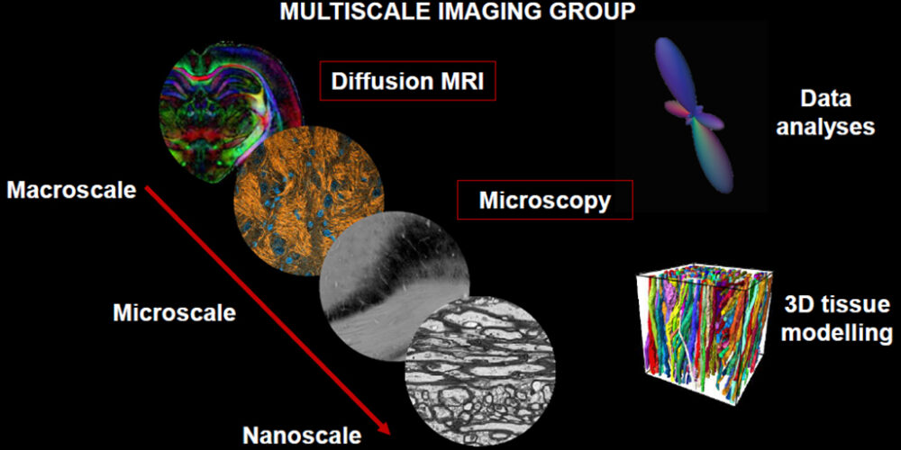
The multiscale imaging group is a multidisciplinary team interested in the application, combination and development of imaging modalities focused in brain at several scales. Our greatest interest is the study of the structure of the healthy and diseased brain from the macro- and microscale with non-invasive techniques to the nanoscale with microscopy techniques in combination with image processing, data analyses and tissue modelling.
Keywords
3D electron microscopy
animal models
biomarkers
brain
brain injury
demyelination
diffusion MRI
diffusion tensor imaging
fourier analysis
high angular resolution diffusion imaging
histology
imaging
inflammation
microimaging
microstructure
montecarlo simulations
neurodegeneration
orientation distribution function
plasticity
segmentation
serial block face scanning electron microscopy
structure tensor
Leaders
Post-doctoral Researchers
-

Jenni Kyyriäinen
Project ResearcherA.I. Virtanen Institute for Molecular Sciences, Faculty of Health Sciences -
Raimo Salo
Project ResearcherA.I. Virtanen Institute for Molecular Sciences, Faculty of Health Sciences -

Juan Valverde Martínez
Visiting ResearcherA.I. Virtanen Institute for Molecular Sciences, Faculty of Health Sciences -
Maxime Yon
Visiting ResearcherA.I. Virtanen Institute for Molecular Sciences, Faculty of Health Sciences
Doctoral Researchers
-

Omar Narvaez Delgado
Doctoral ResearcherA.I. Virtanen Institute for Molecular Sciences, Faculty of Health Sciences -

Melina Estela Dalmau
Doctoral ResearcherA.I. Virtanen Institute for Molecular Sciences, Faculty of Health Sciences -
Mohammad Khateri
Doctoral ResearcherA.I. Virtanen Institute for Molecular Sciences, Faculty of Health Sciences -
Sara Gröhn
Doctoral ResearcherA.I. Virtanen Institute for Molecular Sciences, Faculty of Health Sciences
Technicians
Contact persons
Visiting Scientist
Publications
10 items-
Advanced microscopic validation of diffusion MRI in rat brain
Salo, Raimo. 2021. Publications of the University of Eastern Finland. Dissertations in Health Sciences G5 Doctoral dissertation (article) -
Automated 3D segmentation and morphometry of white matter ultrastructures
Abdollahzadeh, Ali. 2021. Publications of the University of Eastern Finland. Dissertations in Health Sciences G5 Doctoral dissertation (article) -
Cylindrical Shape Decomposition for 3D Segmentation of Tubular Objects
Abdollahzadeh, Ali; Sierra, Alejandra; Tohka, Jussi. 2021. IEEE access. 9: 23979-23995 A1 Journal article (refereed), original research -
DeepACSON automated segmentation of white matter in 3D electron microscopy
Abdollahzadeh, Ali; Belevich, Ilya; Jokitalo, Eija; Sierra, Alejandra; Tohka, Jussi. 2021. Communications biology. 4: 179 A1 Journal article (refereed), original research -
Microstructural Tissue Changes in a Rat Model of Mild Traumatic Brain Injury
Chary Karthik, Narvaez Omar, Salo Raimo A, San Martín Molina Isabel, Tohka Jussi, Aggarwal Manisha, Gröhn Olli, Sierra Alejandra. 2021. Frontiers in neuroscience. 15: 746214 A1 Journal article (refereed), original research -
Assessment of the structural complexity of diffusion MRI voxels using 3D electron microscopy in the rat brain
Salo, Raimo A; Belevich, Ilya; Jokitalo, Eija; Gröhn, Olli; Sierra, Alejandra. 2020. Neuroimage. 225: 117529 A1 Journal article (refereed), original research -
In vivo diffusion tensor imaging in acute and subacute phases of mild traumatic brain injury in rats
San Martín Molina, Isabel; Salo, Raimo A; Abdollahzadeh, Ali; Tohka, Jussi; Gröhn, Olli; Sierra, Alejandra. 2020. eNeuro. 7: ENEURO.0476-19.2020 A1 Journal article (refereed), original research -
Automated 3D Axonal Morphometry of White Matter
Abdollahzadeh, A; Belevich, I; Jokitalo, E; Tohka, J; Sierra, A. 2019. Scientific reports. 9: 6084 A1 Journal article (refereed), original research -
Quantification of anisotropy and orientation in 3D electron microscopy and diffusion tensor imaging in injured rat brain
Salo, Raimo A; Belevich, Ilya; Manninen, Eppu; Jokitalo, Eija; Gröhn, Olli; Sierra, Alejandra. 2018. Neuroimage. 172: 404-414 A1 Journal article (refereed), original research -
Diffusion tensor MRI shows progressive changes in the hippocampus and dentate gyrus after status epilepticus in rat - histological validation with Fourier-based analysis
Salo RA, Miettinen T, Laitinen T, Gröhn O, Sierra A. 2017. Neuroimage. 152: 221-236 A1 Journal article (refereed), original research


