
In vitro and ex vivo electrophysiology core facility

Electrophysiology provides a unique way of investigating cell and tissue functionality. This is important for high quality science not only in basic cell biology and disease investigation but also in the continuously growing fields of stem cells, tissue engineering and therapy development.
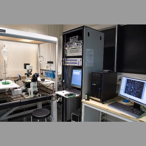
Our in vitro / ex vivo electrophysiology core facility has instrumentation for measurement of single cell electrical and functional properties, including combined fluorescence imaging and patch clamp / intracellular recording systems for cell cultures and tissue slices as well as multielectrode array platform for measurement of neuronal circuits. We provide services on Ca-imaging on cells and slices, patch-clamp experiments, intracellular voltage recordings, cellular and subcellular high speed confocal imaging of ions or voltage, extracellular voltage recordings (cell cultures, small tissues) and MEA measurements of network activities.
We offer personal training, help and guidance for experimental planning, tissue or cell processing and data analysis. We also offer a full service package including experimental design, conduction of the measurements and the following analyses, and provide education in electrophysiology.
Sample type and experiment type are sometimes very research question specific and thus need special protocols to be followed and optimized.
The facility consists of five units:
1- Equipment for patch clamp: The station houses an upright Olympus BX50 microscope equipped with infa red differential interference contrast optics (IR-DIC). Electrophysiological recordings are conducted with a an Axopatch-200B amplifier, optical imaging with a Retiga CMOS imaging camera.Luigs Neumann and Narishige stage controllers regulate sample positioning and electrode movement to the sample Stimulation studies are conducted with a Digitimer DS-3 stimulus isolator unit. Perfusion of thermoregulated fluids and pharmacological investigations in the large bath is achieved by a peristaltic pump and with a fast perfusion system.
Location: Bioteknia 2, 3rd floor, room 3417.
Description: Patch clamp rig for electrophysiological recordings and simultaneous calcium imaging in brain slices or cultured neurons.
Additional info/Comments: use of rig requires operator.
Contact: Tarja Malm, Polina Abushik
2- Equipment for Microelectrode array (MEA) for electrophysiology: PC controlled MEA2100-Mini-System for recording MEAs with 60 electrodes (Multi Channel Systems GmbH), dual automatic temperature controller TC02, Leica SAPO microscope and Leica MC170 HD camera for sample and MEA monitoring during recordings, PPS2 peristaltic pump and temperature controlled PH-01 cannula for delivering drugs to the sample.
Location: Bioteknia 2, 3rd floor, room 3417.
Description: PC controlled MEA system for recording extracellular electrical potentials in tissue slices and cell cultures.
Additional info/Comments: can be used by client after training. Using the drug application system requires an operator. Study planning and analysis requires expert help.
Contact: Tarja Malm, Anssi Pelkonen
3- Equipment for imaging studies in Bioteknia 2, room 3428: TILL Photonics GmbH imaging system including camera and wide spectrum light source based on Olympus IX70 fluorescent microscope platform, patch clamp amplifier, PC controlled rapid solution exchange system, peristaltic pump for delivering drugs to cells, dual automatic temperature controller TC-344B.
Location: Bioteknia 2, 3rd floor, room 3428.
Description: PC controlled imaging system with automatic fast drug application/removal for detection changes in intracellular calcium, reactive oxygen species and mitochondrial membrane potential in primary cells and cell lines.
Additional info/Comments: can be used by client after training.
Contact: Tarja Malm, Rashid Giniatullin, Raisa Giniatullina, Anastasia Shakirzyanova
4- Equipment: Olympus FX1000 confocal microscope combined with micromanipulators, patch clamp amplifier, intracellular voltage amplifier, rapid solution exchange system, perfusion system for cells and tissues, microinjection, temperature and gas control and feedback controlled objective heaters.
Location: Bioteknia 1, 4th floor, room 4103.
Description: Fully equipped set-up for simultaneous live cell/tissue confocal imaging and electrophysiology.
Additional info/Comments: Requires operator.
Contact: Pasi Tavi, Nikolay Naumenko
Instrument usage:
Ca-imaging unit: 26€/hour
Total hourly rate for university internal projects (from 16.00 to 08:00), cheaper evening and weekend pricing reduces the workload of day hours by 15€/hour.
Sales price to outsiders (incl. VAT 24%, and 24% overhead, 20% margin) 44€/hour Consultation price for outsiders (incl. VAT 24%, and 24% overhead, 20% margin, indirect employee costs 45%) 62€/hour.
Multichannel systems MEA: 10€ /hour
Sales price to outsiders 10€/hour (incl. VAT 24%, and 24% overhead, 20% margin) 19€/hour.
Consultation price for outsiders (incl. VAT 24%, and 24% overhead, 20% margin, indirect employee costs 45%) 62€/ hour.
Cooperation
-
 Neuroinflammation research group 01.01.2017 -
 -
 Molecular Physiology 01.01.2009 -
 -
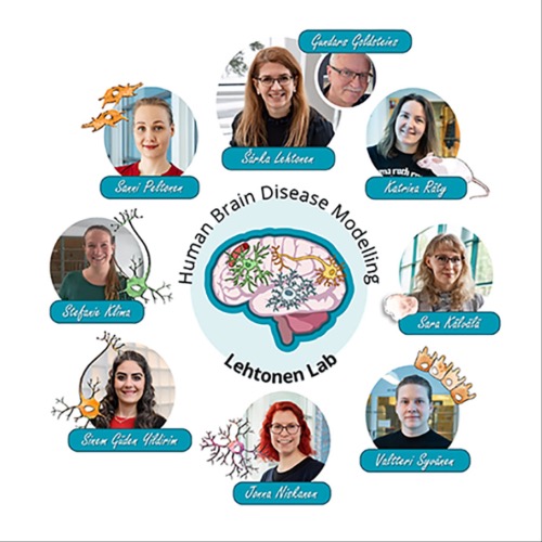 Human Brain Disease Modelling (Lehtonen lab) 01.01.2021 -
 -
 Cancer Cell Plasticity - Ketola Lab 01.09.2019 -
 -
 AIVI Stem Cell Center
 -
 Annakaisa Haapasalo Group (Molecular Neurodegeneration) 01.06.2015 -
Keywords
Members
-

Tarja Malm
ProfessorA.I. Virtanen Institute for Molecular Sciences, Faculty of Health Sciences -
Pasi Tavi
ProfessorA.I. Virtanen Institute for Molecular Sciences, Faculty of Health Sciences -
Rashid Giniatullin
Research DirectorA.I. Virtanen Institute for Molecular Sciences, Faculty of Health Sciences -
Anssi Pelkonen
Staff ScientistA.I. Virtanen Institute for Molecular Sciences, Faculty of Health Sciences -

Anastasia Shakirzyanova
Postdoctoral ResearcherA.I. Virtanen Institute for Molecular Sciences, Faculty of Health Sciences -
Raisa Giniatullina
Project ResearcherA.I. Virtanen Institute for Molecular Sciences, Faculty of Health Sciences -
Nikolay Naumenko
Project ResearcherA.I. Virtanen Institute for Molecular Sciences, Faculty of Health Sciences