Cardiac imaging group
Leaders
Magnetic resonance imaging (MRI) is a non-invasive and radiation-free medical imaging modality, whose strength is its high soft tissue contrast with high spatial and temporal resolution without subjecting the patient to ionizing radiation. There are numerous ways to generate good contrast in an MR image. These ways are divided mainly into endogenous and exogenous contrasts. MRI is a versatile tool for imaging the heart since it can provide anatomical, functional and pathological information of the myocardial tissue.
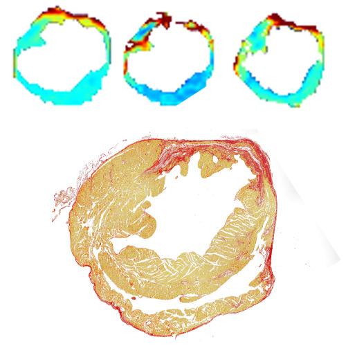
Cardiovascular diseases (CVD) and ischemic heart diseases are the leading global causes of death. There are many CVD, for example myocardial infarction and cardiac inflammation with their sub-diseases. CVD are affecting to the myocardium by damaging it and accumulation of fibrotic tissue, which are causing the decrease of heart function. It is important to image non-invasively the earliest phases of different CVD and make the cardiac imaging more sensitive to diagnostic in different CVD as earlt as possible. Then the characteristic development of each CVD would be able to image non-invasively.
Projects of this research group is to develop novel and more sensitive imaging methods for the diagnostic of different CVD. For example in MRI, new methods to excite and collecting the signal are the ways to develop these kind of methods.
Cooperation
Projects
Preclinical:
-Novel methods to excite and collecting the MR-signal
-PET-MRI development for cardiac imaging
-Modern methods to cardiac MRI
Clinical:
-MOMESY-study
-CoHeDI MRI-study
-DoCS-MRI-study


